 |
| This article contains graphic images from real cases which may be disturbing to some viewers. Viewer discretion is advised. |
A head injury is any trauma to the scalp, skull, or brain. The injury may be only a minor bump
on the skull or a serious brain
injury. Head injury can
be either closed or open (penetrating). A closed head injury means you received a hard blow to the head from striking an object, but the
object did not break the skull. The head injuries are the most common ones, and
at the same time, the most dangerous. Innocently looking trauma may lead to a
demise, though, a very severe looking
one may be come away unscathed from (a blow/ accident). It all lies in a
sheer luck, sometimes.
The
head and brain trauma might be categorized according to the mechanism it is
done:
-
impact injuries;
-
acceleration injuries; declaration
injuries which divides into subdural hematomas and diffuse axonal injuries.
The
impact injuries are caused when an object strikes or is struck by the head.
These injuries consist of the local effects of contact between the head and the
object and these injuries are:
-
soft
tissue injuries which include lacerations, abrasions, contusions of the scalp;
-
fracture
of the skull;
-
contusions
of the brain;
-
intracerebral
hemorrhages;
Acceleraction
or declaration injuries emerge due to sudden movement of the head the instant
after the injury, the outcome of that movement is intracranial pressure during
which the brain is exposed to shearing and tensile forces.
Subdural
hematomas are secondary to tearing of the subdural bridging veins; diffuse
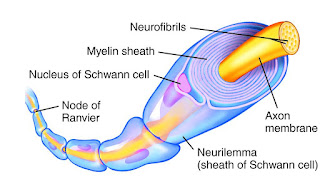
axonal injury is secondary to injury to the [1]axons.
While acceleration or deceleration injuries are associated with impact which is
not necessary for the production of these injuries, just sudden angular
rotation of the head. In situations encountered by forensic pathologists,
however, acceleration or deceleration injuries of the brain involve impact.
To
illustrate more the seriousness of the head and brain trauma all injuries will
be described and analysed, first will be impact injuries; soft tissue injuries.
Soft tissue injuries occur when the head is struck by an object or strikes the
ground, the initial injuries are incurred by the scalp — lacerations,
contusions, or abrasions. Lacerations produce profuse bleeding due to the great
vascularity of the scalp. Only under the most unusual circumstances would this
be life threatening. More serious traumas are fractures of the skull this is the
second type of injury that can be incurred is to the skull. In general, whenever
a head is either struck with or strikes an object having a broad flat surface
area, the skull at the point of impact flattens out to conform to the shape of
the surface against which it impacts. As the skull is flattened and bent
inward, adjacent, but more distant areas, are bent outward by a wave of
deformation consisting of the central area of inbending and the peripheral outbending.
The outbending can occur at a considerable distance from the point of impact.
Where the skull curves sharply, the extent of inbending and outbending is not
so great as in less-curved areas. If a fracture of the skull occurs, the
fracture does not begin at the point of impact, but at the point of outbending.
Linear fractures begin on the external surface of the skull by the forces
produced by the outbending of the bone. After inbending, the skull attempts to return
to its normal configuration. As the inbent portion of the skull does so, the
fracture line extends from its originating site toward the area of impact, as
well as in the opposite direction. The fracture line may or may not reach the
point of impact and could actually continue through it.
The head
deformation and the impact of the disfigurement depends on the following
factors:
-
the
amount of hair;
-
the
thickness of the scalp;
-
the
thickness of the skull;
-
the
elasticity of the bone at the point of the impact;
-
the
shape, weight, and consistency of the object impacted or impacting by the head;
-
the
velocity at which either the blow was delivered or the head strikes the object.
The
amount of energy required for production of a single linear fracture from a
low-velocity blow or fall depends on whether the head strikes a hard unyielding
surface or a relatively soft yielding surface. With a yielding surface, a large
proportion of the impacting energy is transferred to the surface by way of the
deformation of the surface, thus decreasing the amount of energy available to
cause head injury. In the case of a hard unyielding surface, e.g., a steel
plate there is essentially no energy transferred to the impacted surface. If a
head strikes or is struck by a deformable object, not all the energy possessed
by either the object or head will be available for deformation of the skull. At
impact, the object will tend to indent and deform so as to wrap itself around
the head. Thus, the energy delivered is no longer in a localized focus but is
dispersed over a considerable area, reducing the possibility of a skull
fracture. Linear or comminuted fractures of the skull produced by impaction of
a head and a relatively soft and flexible object, such as the instrument panel
of a motor vehicle, require kinetic energy levels at impact of between 268 and 581 ft lbs. Simple linear fractures are typically seen in low-velocity
impacts with a large area of contact between the head and impacting object. A
fall to the pavement is the best example. With increased velocity and, thus,
greater force, one may have a series of complete or incomplete circular fractures encircling the impact point; These
fractures result from failure of the external surface of the bone at the edge
of the inbent area, due to extreme inbending at the time of impact. If the
velocity and energy of impact are increased even more, one gets stellate fractures, where there is depression of the
bone at the point of impact. The severe inbending about the impact site
produces fractures on the inner surface that radiate out from the site of the
blow. Fractures resulting from the outbending of the bone at a distance from
the point of impact, and arising in the outer surface of the skull, extend toward
the point of impact and join with the fractures radiating outward from the
point of impact. Circular fractures may occur at the junction of the inbending
bone on its external surface.
A depressed skull fracture occurs when the skull is struck with
an object having a relatively large amount of kinetic energy but a small
surface area when an object with a large amount of kinetic energy impacts only
a small area of the skull. The scalp does not significantly affect the nature
of the injuries to the skull. Large deformations occurring at a distance from
the point of impact are no longer present. At the point of impact, there is a depressed
fracture, possibly with fragmentation. The fractures are due to failure of the
inner surface of the skull secondary to the inbending. An example of this type
of fracture is the circular depressed fracture of a hammer blow.
Blows in different areas of the head can have
different effects for example blow to the top of the head tends to produce a cranial vault fracture
that might or might not extend into the temporal region or base of the skull. Basilar skull fractures are
quite common in forensic medicine. The base of the skull, by virtue of its
construction and irregular shape, is weak. Almost any diffuse impact to the
vertex of the skull will produce basilar fractures. Basal skull fractures can
occur from blows anywhere along the circumference of the skull below the
cranial vault. They can run anterior-posteriorly, posterior- anteriorly, side
to side and any combination of these three. Basal skull fractures may be missed
on X-rays of the skull. With a basal fracture, intracranial passage of a
nasogastric tube or nasophrayngeal airway can occur. Hinge fractures are transverse fractures of the base
of the skull that completely bisect the base of the skull, creating a “hinge.” The fractures are divided into three categories:
- coronal plane fractures;
- the fractures which run from front to the contralateral back,
passing through the sella turcica;
- the fractures which run from side to side in the coronal plane but do not pass through the sella turcica.
Impact
injuries can produce contusions and lacerations of the brain. Contusions are
the most frequently encountered traumatic lesion of the brain. Contusions involve
the crests of the gyri, but can extend into the white matter as wedge shaped
lesions. Cortical contusions consist of areas of hemorrhage and necrosis that
originate at the moment of impact. The amount of bleeding depends on the type
(small artery, capillary, vein) and caliber of the vessel injured as well as if
there is any adjacent necrosis. If necrosis is present, the contusions develop
a wedge-shaped appearance with the base of the wedge at the crest and the point
toward the white matter. If there is profuse bleeding, the areas of hemorrhage
may expand into the white matter or subarachnoid space, essentially forming an
area of intracerebral hemorrhage.
(streak-like linear contusion)
Intracerebral hematomas are discrete collections of blood within the cerebral parenchyma that
are not in contact with the surface of the brain. They are principally located
in the white matter of the fronto-temporal lobes and are caused by impact. They
are said to differ from hemorrhagic intermediary coup contusions in that they
are well demarcated, homogeneous collections of blood, in contrast to
contusions that are blood and contused cerebral parenchyma. In the authors’
opinion, this distinction is artifactual may reflect a longer survival time by
the intracerebral hematomas with continued bleeding. One of the most interesting
aspects of intracerebral hematomas is that they can appear hours to days after
the injury. There have been numerous cases where a computerized tomography (CT)
scan on admission has shown no intracerebral hematomas, but subsequent CT
scans, taken several hours to several days after admission, revealed some. In
some instances, their development has been followed by successive CT scans. Primary
intracerebral hemorrhages involving the basal ganglia which are found in approximately
10% of fatal head injuries. They are caused by deceleration/ acceleration
forces and are commonly found in association with diffuse axonal injury and
gliding contusions. In 90% of the cases, the cause of the injury was either a
motor vehicle accident or a fall.
Some
individuals do not develop significant symptoms of a subdural hematoma for
weeks to months after head injury. The resultant hematoma is a chronic subdural hematoma. In this
entity, instead of the initial acute subdural hematoma’s becoming organized and
then smaller as it is reabsorbed, it begins to enlarge. This continues until
the chronic subdural hematoma produces sufficient symptomology. The victims of
chronic subdural hematomas tend to be either infants younger than 6 months of
age or the elderly.
Any brain
injury leaves irremovable traces, the scope of it depends on the medical
treatment One has been given, the scope and the type of injury itself, the tool
that has been used, believe to understand, no matter how slight, small, tiny,
and irrelevant the trauma may look like, it has to be checked – accidental or
none-accidental, do not be afraid to report it, it can take months to recall itself and at that point, it can kill.
Bibliography:
1. Criminal Investigations – Crime Scene Investigation.2000
2. Forensic Science.2006
3. Techniques of Crime Scene Investigation.2012
4. Forensics Pathology.2001
5. Pathology.2005
6. Forensic DNA Technology (Lewis Publishers,New York, 1991).
7. The Examination and Typing of Bloodstains in the Crime Laboratory (U.S. Department of Justice, Washington, D.C., 1971).
8. „A Short History of the Polymerase Chain Reaction". PCR Protocols. Methods in Molecular Biology.
9. Molecular Cloning: A Laboratory Manual (3rd ed.). Cold Spring Harbor N.Y. Cold Spring Harbor
10. "Antibodies as Thermolabile Switches: High Temperature Triggering for the Polymerase Chain Reaction". Bio/Technology.1994
11. Forensic Science Handbook, vol. III (Regents/Prentice Hall, Englewood Cliffs, NJ, 1993).
12. "Thermostable DNA Polymerases for a Wide Spectrum of Applications: Comparison of a Robust Hybrid TopoTaq to other enzymes". In Kieleczawa J. DNA Sequencing II: Optimizing Preparation and Cleanup. Jones and Bartlett. 2006
13. "Microscale chaotic advection enables robust convective DNA replication.". Analytical Chemistry. 2013
14. Sourcebook in Forensic Serology, Immunology, and Biochemistry (U.S. Department of Justice, National Institute of Justice, Washington, D.C.,1983).
15. C. A. Villee et al., Biology (Saunders College Publishing, Philadelphia, 2nd ed.,1989).
16. Molecular Biology of the Gene (Benjamin/Cummings Publishing Company, Menlo Park, CA, 4th ed., 1987).
17. Molecular Evolutionary Genetics (Plenum Press, New York,1985).
18. Human Physiology. An Integrate. 2016
Acknowledgements:
The Police Department;
https://www.politie.nl/mijnbuurt/politiebureaus/05/burgwallen.html and a Chief Inspector – Mr. Erik Akerboom ©
[1] An axon is
a long, slender projection of a nerve cell, or neuron, that typically
conducts electrical impulses away from the neuron's cell body.
Axons are also known as nerve fibers. The function of the axon is to
transmit information to different neurons, muscles and glands. In certain
sensory neurons (pseudounipolar neurons), such as those for touch and warmth,
the electrical impulse travels along an axon from the periphery to the cell
body, and from the cell body to the spinal cord along another branch of the
same axon. Axon dysfunction has caused many inherited and acquired neurological
disorders which can affect both the peripheral and central neurons. Nerve
fibers are classed into three types – A delta fibers, B fibers, and C
fibres. A and B are myelinated and C are unmyelinated.An axon is one of
two types of protoplasmic protrusions that extrude from the cell body
of a neuron, the other type being dendrites. Axons are distinguished from
dendrites by several features, including shape (dendrites often taper while
axons usually maintain a constant radius), length (dendrites are restricted to
a small region around the cell body while axons can be much longer), and
function (dendrites usually receive signals while axons usually transmit them).
Axons are covered by a membrane known as axolemma: the cytoplasm of axon
is called axoplasm. The branched end of an axon is formed
by telodendria: the swollen end of a telodendron is known as the axon
terminal; which joins the dendron or cell body of another neurone forming a
synaptic connection. Some types of neurons have no axon and transmit signals
from their dendrites. No neuron ever has more than one axon; however in
invertebrates such as insects or leeches the axon sometimes consists of several
regions that function more or less independently of each other. Most axons branch, in some cases very
profusely. Axons make contact with other cells—usually other neurons but
sometimes muscle or gland cells—at junctions called synapses. At a
synapse, the membrane of the axon closely adjoins the membrane of the
target cell, and special molecular structures serve to transmit electrical or
electrochemical signals across the gap. Some synaptic junctions appear partway
along an axon as it extends—these are called en passant ("in
passing") synapses. Other synapses appear as terminals at the ends of
axonal branches. A single axon, with all its branches taken together,
can innervate multiple parts of the brain and generate thousands of
synaptic terminals.


Komentarze
Prześlij komentarz